
The Medical Lens: Highlights from the Stanley B. Burns, MD, Historic Medical Photography Collection
January 27, 2023 - March 10, 2023 (EXTENDED TO MARCH 19TH!)
Join us at the Medical Library for our newest Rotunda exhibition! Medicine is a field grounded in the visual world. Over the centuries, illustration became increasingly embedded in the medical field via textbooks, posters, and other visual medium. With the development of photography in the early nineteenth century, medicine acquired a new way of viewing the patient. Besides being integrated in medical education and training, photography became a means of creating professional identity. To the larger world, medical photography helped shape the image of medical care and the profession, promoted technological advancements, sold products, and influenced public policy.
The Medical Lens explores the importance of photography in medicine through images selected from the recently acquired Stanley B. Burns, MD, Historic Medical Photography Collection at Yale University. The collection encompasses a wide variety of photographic and print techniques including daguerreotypes, ambrotypes, and tintypes from the earliest years of photography, cartes de visite, cabinet cards, lantern slides, photo albums and collections of prints assembled by medical practitioners, postcards, and publications.
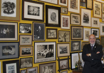
Stanley B. Burns, MD, FACS, is an ophthalmologist and Research Professor of Medicine and Psychiatry, and Professor of Medical Humanities at New York University: Langone Health. He began collecting historic photography in 1975, and over time amassed over a million images that he curated in multiple books, articles, and exhibitions. Dr. Burns is pictured standing in front of his photo wall containing some of the most iconic images from the Burns Archive, which he established in 1977.
This exhibition is curated by Katherine Isham, MLIS, and Melissa Grafe, PhD, with the valued expertise of Stanley B. Burns, MD, FACS. The curators want to thank Chris Zollo, Kelly Perry, Laura O’Brien-Miller, Terry Dagradi, Dana Haugh, and Melanie Norton for their additional assistance in bringing this exhibition to life.
Please see the exhibition object list to begin exploring the items on display.
Click to open the object list
Case 1: Introduction
-F. R. Reynolds and classmate before and after receiving their medical degrees at Rush Medical College, tintypes, 1883
-Florence Nightingale photographed by H. Hering, “Photographer to the Queen,” carte de visite, circa 1856-1857
-James Samuel How (Howe), MD, dead from cholera epidemic, St. Louis, Missouri, daguerreotype with obituary notice, 1849
-Ava V. Chadwick-Herns’s Battle Creek Sanitarium pamphlet with added photographs and notations, Battle Creek, Michigan, 1906-1907
-“Synoviales de la main” (dissection of the hand to show synovial system), 1870 and “Pelvi-support contre-extenseur” (counter-tension pelvic support), 1873 from Revue Photographique des hôpitaux de Paris. Gift of Stanley B. Burns, MD, 2020.
Case 2: Medical Identity and the Profession
(1) Meade brothers studying medicine, Victor, New York, tintype, circa 1860-1865
To be replaced mid-February with:
(1) Two medical students studying anatomy with book, bones, and dissected arm, tintype, circa 1860-1865
(2) Dental extraction staged scene, tintype circa 1855-1865
(3) Portrait of Dr. Matthew Gill, “A student of Esculapius,” photographer S. Krausz, Chicago, cabinet card, circa 1891-1892
(4) Portrait of a visiting nurse, photographer John Suchy, Chicago, cabinet card, circa 1898-1900
(5) “Dr. Gridley’s first operation,” amputation surgery staged in a photography studio, photographer W. A. Hopkins & Company, Rapid City, South Dakota, cabinet card, 1891
(6) Elderly pharmacist with bottles of medicines, hand-tinted ambrotype, circa 1860-1866
(7) Physician/pharmacist using microscope, New York, gelatin silver print, circa 1895
(8) Pharmacist and assistant in a pharmacy, gelatin silver print, circa 1900
(9) Portrait of Danish nurse with red cross armband, photographer Mary Steen, Copenhagen, carte de visite, circa 1893
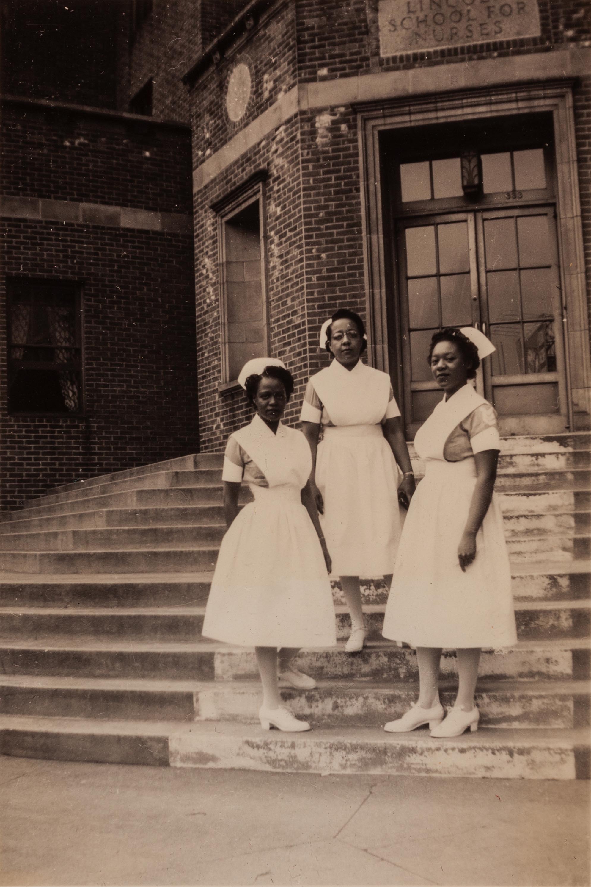
(10) Three nurses on the steps of the Lincoln School for Nurses, Bronx, New York, gelatin silver print, circa 1930
(11) Fordham Hospital medics with horse-drawn ambulance, gelatin silver print, circa 1892-1900
(12) Group photo of women interns at the Children’s Hospital of San Francisco, gelatin silver print, 1925-1926
Case 3: Medical Spaces and their Meanings
(1) Dr. Bernstein, dentist, in his office, gelatin silver print, circa 1945
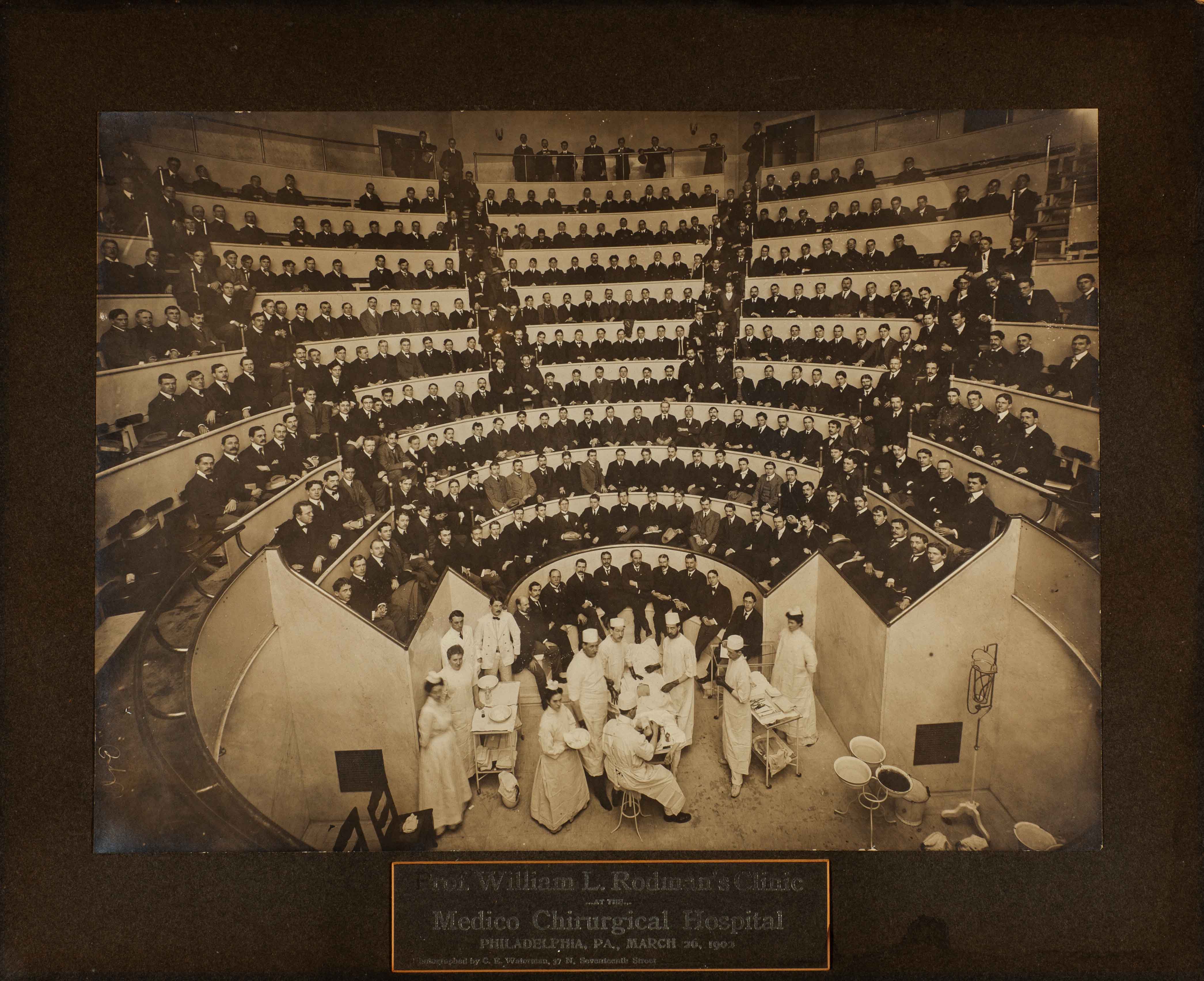
(2) Surgeon William L. Rodman’s clinic in the operating theater of the Medico Chirurgical Hospital, Philadelphia, photographer C. E. Waterman, gelatin silver print, March 26, 1902
(3) Exterior view of Mount Sinai Hospital from series “Views in New York City and Vicinity,” stereoview card, 1893
(4) Operation taking place in a Bellevue ward circa 1880s-1890s, gelatin silver copy print, 1948
(5) Operation led by female surgeon, gelatin silver print, circa 1905-1920
(6) Receiving wards, from George Pfaler E.M.D.’s Old Blockley Hospital photo album, Philadelphia, gelatin silver print, 1900-1901
(7) Boston City Hospital Ward P, gelatin silver print, Christmas 1912
Case 4: The Boom of Medical Innovation and Technology
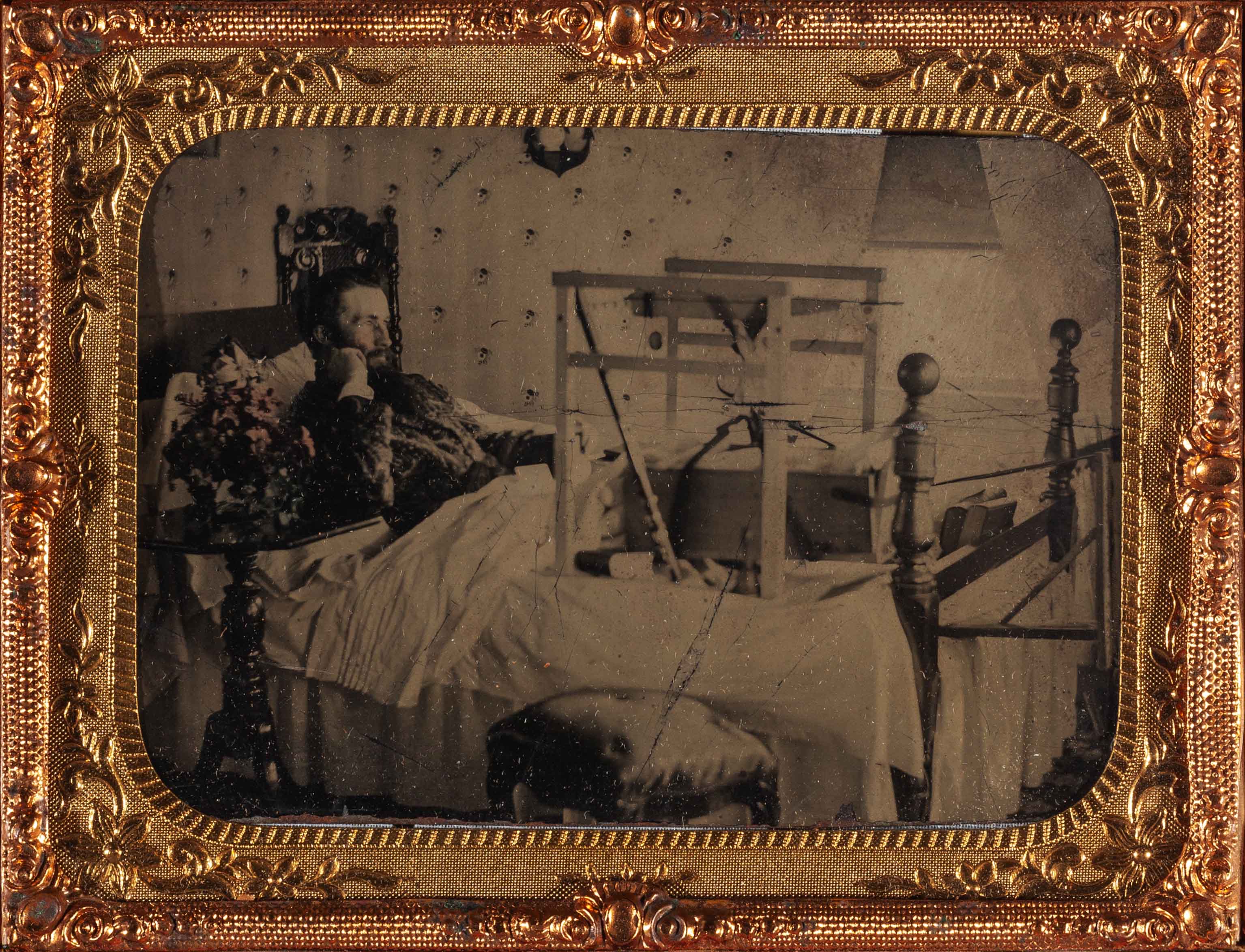
(1) Man in bed with leg in an early traction device, tintype in thermoplastic case, circa 1860-1870
(2) “Artificial sunlight for children,” showing a child receiving a “light bath” treatment at New York Nursery and Child’s Hospital, Keystone View Company, Inc., gelatin silver print, circa 1920-1935
(3) “Making ‘movies’ of the heart,” Kymograph machine combining X-ray and moving picture technology built by Dr. Wendell G. Scott and Dr. Sherwood Moore of Washington School of Medicine in St. Louis, International News Photo, gelatin silver print, 1936
(4) “New electron microscope has great range,” Dr. Gordon Scott of Washington University Medical School using an electron microscope, Acme Chicago Bureau, gelatin silver print, 1940
(5) “Machine will act as heart or lung,” created by J. Jongbloed of Holland for use during surgery, shown at conference of surgeons at the Sorbonne, Paris International News Photos, gelatin silver print, 1951
(6) “Skin resistance to sun measured,” Dr. Robert C. Burt of Pasadena, CA demonstrating his device for measuring how long one may be exposed to sunlight without injury, gelatin silver print, circa 1920-1930
(7) “La formule ideale de sang artificiel” (the ideal formula for artificial blood), Dr. Gottendenker of Vienna with his new invention: artificial human blood, Agence Trampus, gelatin silver print, 1937
(8) The “Headshrinker” positron detector invented by James S. Robertson at Brookhaven National Laboratory, a direct forerunner of positron emission tomography scanning, photographer unknown, gelatin silver print, 1961
(9) “Une nouvelle methode de traitement pour le cancere” (a new way to treat cancer), radiation sphere invented by Anton Zeeman and Doctor Erwin Fuhrer for the treatment of cancer, Agence Trampus, gelatin silver print, 1938
Case 5: Diseases, Vaccines, and Treatments
(1) Child with smallpox, New York City, gelatin silver print, 1881
(2) Scenes from pneumonic plague in China, gelatin silver prints, 1911.
Pictured are four doctors with thick face masks standing in front of a train; a doctor being sprayed with disinfectant; a doctor and medical assistants with horse-drawn carts for living and dead plague victims; and a doctor and military personnel standing outside an infected building that’s being burned down to stop the spread of disease.
(3) Hookworm Disease Commission in Jamaica, gelatin silver prints, circa 1918
In these images from a larger album, medical personnel are using microscopes to examine samples and encouraging local people to see the hookworm eggs under the microscope as part of a health demonstration.
(4) Elizabeth Kenny demonstrating physical therapy treatment on a young polio patient for nurses at General Hospital, Minneapolis, gelatin silver print, circa 1940
(5) Female scientists preparing vaccines in the Pasteur Institute toxins and antitoxins department, Photograph Trampus, Paris, gelatin silver print, 1943
(6) Adding formalin to transform toxin into antitoxin at the Pasteur Institute, Photograph Trampus, Paris, gelatin silver print, 1943
(7) U.S. Army Captain Daniel Staples administering typhoid vaccine to a young refugee from a flood area, Forrest City, Arkansas, International Newsreel, gelatin silver print, 1927
(8) Man being vaccinated at Pasteur Institute, photo postcard published by Neurdein et Cie, Paris, 1916
(9) Catholic missionary staff administering vaccines, photo postcard published by La Propagation de la Foi, Paris/Lyon, circa 1920
Case 6: War and Medicine
(1) Civil War contract surgeon in his tent with books, medications, and medical bag, tintype, circa 1862-1865
(2) Surgical scene in front of a tent at Camp Letterman, Gettysburg, partial stereoview card, July 1863. Gift of Stanley B. Burns, MD, 2022
(3) American Women’s Hospital ambulance driver with her vehicle, photographer E. Belval, France, gelatin silver print, circa 1918
(4) World War I military doctor treating soldier with leg wound in multi-patient clinic, gelatin silver print, circa 1914-1918
(5) Back view of World War I soldier with severe chest injury recovering at Walter Reed Hospital, gelatin silver print, circa 1917-1920
(6) Wounded soldiers posing after recovery with wax models of their facial wounds from Kriegszahnklinik der IV. Armee in Lublin, a German army maxillofacial surgery album, 1916
(7) French World War I veteran photographed with his leg prosthesis from Considérations sur la Rééducation Professionelle Dans les Industries du Bâtiment (Considerations on vocational retraining in the construction industries), one of the first state-funded veteran rehabilitation programs, Charles Vallee, MD, France, 1917
(8) World War II medics administering plasma to battle casualty “on the run” to an L-5 plane for evacuation, Mindanao, Philippines, U.S. Army photograph, gelatin silver print, circa 1941-1942
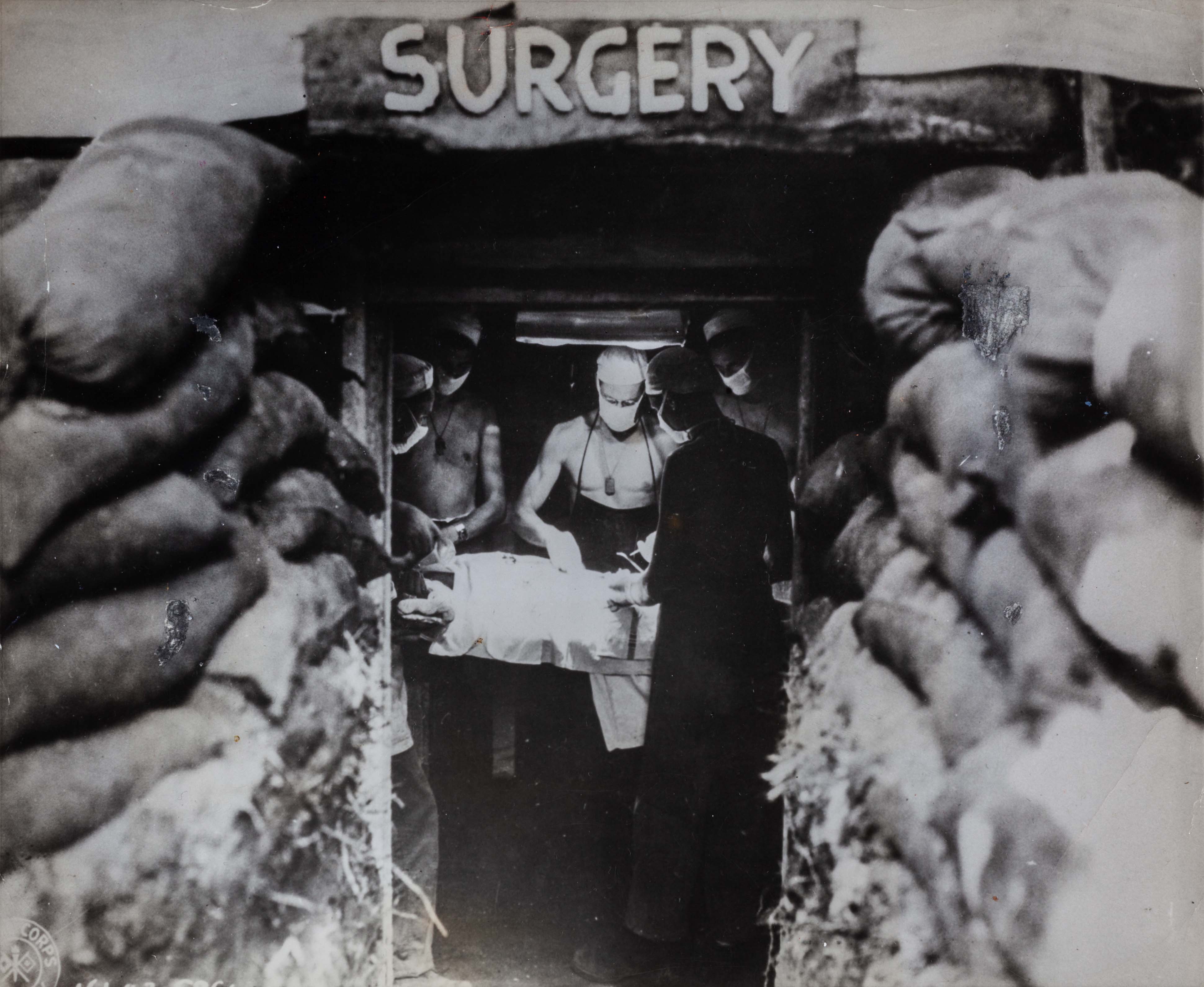
(9) American Army surgeon operating on wounded soldier in underground surgery, Bougainville, Papua New Guinea, U.S. Army photograph, gelatin silver print, 1943
Case 7: Patient Photography and Diagnostics
(1) Civil war veteran receiving morphine injection from a physician, photographer B. Perry, Chamberlain, South Dakota, cabinet card, circa 1865-1866
(2) Nurse taking the pulse of female patient in a wheelchair, photographed by Altman and Edelman, Battle Creek, Michigan, cabinet card, circa 1894-1895
(3) Portrait of an obese man with edema of leg, tintype, circa 1865-1875
(4) Portrait of man with facial and neck tumor, photographer J. G. Ellinwood, Manchester, New Hampshire, carte de visite, circa 1871-1910
(5) Photograph documenting the spinal alignment of a young woman from Berkeley Gymnasium log book on student posture, photographer M. K. Wallin, MD, gelatin silver print, circa 1904-1909
(6) “Tubercular sylphide (on a woman’s back). From the collection of photographs of skin diseases of Dr. George Henry Fox,” page from The Medical Record: Weekly Journal of Medicine and Surgery, December 31, 1887
(7) Man with carcinoma of neck before and after treatment and with his family, Allentown, Pennsylvania, gelatin silver prints attached with surgical tape, circa 1915
(8) Lantern slides of a woman with fractured arm: x-rays and with her arm splinted, circa 1920-1930
(9) “Dr. Bordiu, marquis of Villa Verde, studies X rays during operation performed on Spanish child born with heart ailment,” photographer Jose Maria Lara, Pix Incorporated, New York City, gelatin silver print, circa 1950-1969
(10) Microscopic photography by Carlos Finlay, MD, from his research on yellow fever in Havana, Cuba: “Yellow fever blood, first day, fatal case x1450” and “Yellow fever blood, 5th day, fatal case x1450,” cabinet cards, 1879
Case 8: Teaching Medicine
-Cartes de visite documenting Civil War veterans’ wounds and recovery, compiled by Dr. Reed Bontecou, Surgeon-in-Charge of Harewood U.S. Army General Hospital, Washington DC, circa 1863-1864, and donated to Army Medical Museum. Gift of Stanley B. Burns, MD, 2022.
AND
-Annotated teaching prints of injured Civil War soldiers: James Middleton with gunshot wound through the left shoulder and unidentified soldier with wound on left thigh, Dr. Reed Bontecou, enlargements of albumin prints, circa 1864-1865. Gift of Stanley B. Burns, MD, 2020.
-Stereo prints from Lernt helfen (Learn to help), a 3D first aid guide for lay helpers that was packaged with a small folding stereoscope viewer, Germany, 1952
-Lantern slides created by Dr. Cutler using pre-made mats from William Garrison Reed, Boston, circa 1890
- On view are slides on “Purpurra haemorrhagica on leg” and “Herpes zoster on eye.”
-To be replaced mid-February with slides on “Purpura rheumatica” and “Tinea Versicolor.”
-“Tying the artery after the anastomosis is made” stereoview photograph from Transfusion of Blood by G. W. Crile, from Howard Kelly’s Stereo-clinic series, 1913
AND
-“Closing the wound. Drainage.” stereoview photograph from Thyroidectomy for Exophthalmic Goiter by A. H. Ferguson, from Howard Kelly’s Stereo-clinic series, 1911. With stereoscope, circa 1890-1915
Opening Tour and Special Program:
Thursday, February 9th
4:15pm – 4:45pm - Meet the curators and Dr. Burns and explore The Medical Lens through a short opening tour. Light refreshments will be served. Cushing Rotunda, Cushing/Whitney Medical Library
5pm – 6pm - “Medical Photography and the Humanities: Connecting History to Practice,” a session with Stanley B. Burns, MD, FACS and Chitra Ramalingam, PhD. Co-sponsored by The Program for Humanities in Medicine at Yale School of Medicine. Room 115, just off the Cushing Rotunda, Cushing/Whitney Medical Library. The recording of the session is now available online through The Program for Humanities in Medicine website.
Stay tuned for additional tour announcements for this limited time exhibition!